Teaching in courses:
Research Group
Neurophysics

Group leader: Axel Thielscher
The Neurophysics group is headed by Professor Axel Thielscher and situated both at DRCMR and DTU Health Tech. We work on advancing non-invasive transcranial brain stimulation to modulate human brain activity and open up new therapeutic possibilities. Our aim is to develop and apply multi-scale computational methods to decipher the involved physical, biophysical and physiological mechanisms of action that give rise to the observed physiological and clinical stimulation effects. We apply this knowledge to optimize and personalize the stimulation, with the aim to contribute to new therapeutic approaches.
We complement and validate our computational research on neurostimulation by neuroimaging such as functional MRI and electroencephalography to characterize the in-vivo impact of neurostimulation on brain activity, levering work both within the group and in national and international collaborations.
Selected publications:
Thielscher A, Antunes A and Saturnino GB (2015), Field modeling for transcranial magnetic stimulation: a useful tool to understand the physiological effects of TMS? IEEE EMBS 2015, Milano, Italy
Main paper introducing SimNIBS, > 900 citations on Google scholar
Saturnino GB, Siebner HR, Thielscher A*, Madsen KH* (2019) Accessibility of cortical regions to focal TES: Dependence on spatial position, safety, and practical constraints. Neuroimage 203, 116183
* shared last authorship
Research focus
SimNIBS
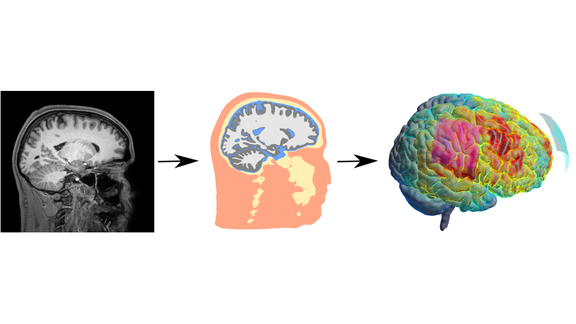
SimNIBS is a scientific, open-source software project which enables the user to simulate electric fields in the brain induced by transcranial brain stimulation (TBS). Accurate simulations require realistic models of the individual head anatomy, knowledge of tissue properties like conductivity, and, optionally, predictions of how neurons respond to externally applied electric fields. We also work on methods for optimizing coil positions for magnetic TBS and electrode configurations in electrical TBS. SimNIBS combines the research from most of our projects and includes important contributions from international collaborators. It makes the methods which we develop accessible to the research community.
Website: www.simnibs.org
Transcranial Ultrasound Stimulation

Transcranial Ultrasound Stimulation (TUS) is a promising new technology to non-invasively modulate neuronal activity in the brain. The key benefit of ultrasound stimulation compared to magnetic and electric stimulation is a much smaller stimulation target area. One of the challenges currently facing TUS is precisely focusing the beam onto the target region through the skull. We work on developing and validating deep learning-based methods to predict head anatomy and tissue properties from magnetic resonance images. These properties are used in acoustic simulations of ultrasound and can improve the efficacy and safety of ultrasound stimulation.
People:
Collaborators:
Characterizing the Neural Response to Non-Invasive Brain Stimulation

This project examines how transcranial magnetic and electric stimulation activate neurons in the human brain. The focus is on understanding the underlying biophysical mechanisms and on applying this knowledge to determine how technical parameters and interindividual differences affect the stimulation effects. The collaborative NeuroSimNIBS project creates new computational tools that translate our findings to clinical research on new treatment protocols with improved precision and efficacy.
People:
Collaborators:
Brain and Head segmentation
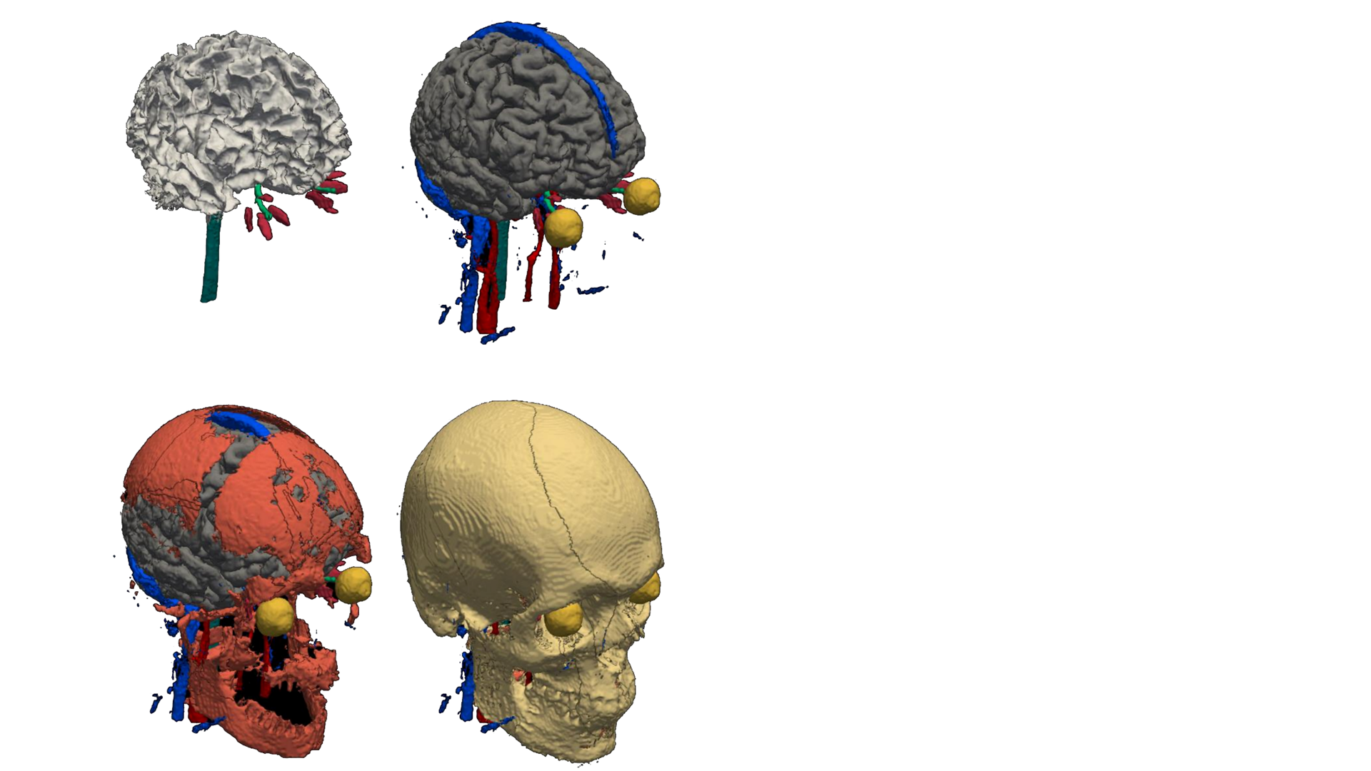
The head and brain anatomy, and microstructural tissue properties vary from individual to individual. These variations have a large effect on the physics and biophysics of brain stimulation. For example, they affect how electric currents produced by non-invasive brain stimulation propagate through the head and brain, and how neurons react to the induced currents.
We develop tools to automatically segment the head and brain structures of a person from his/her MRI scan. The segmentation can, e.g. be used to inform individually targeted brain stimulation or to precisely localize brain activity using source localization. A key focus for our group is to ensure that the tools work robustly across different MRI scanners and scan protocols.
People:
Collaborators:
Measuring current flow and physiological effects of TBS in the human brain
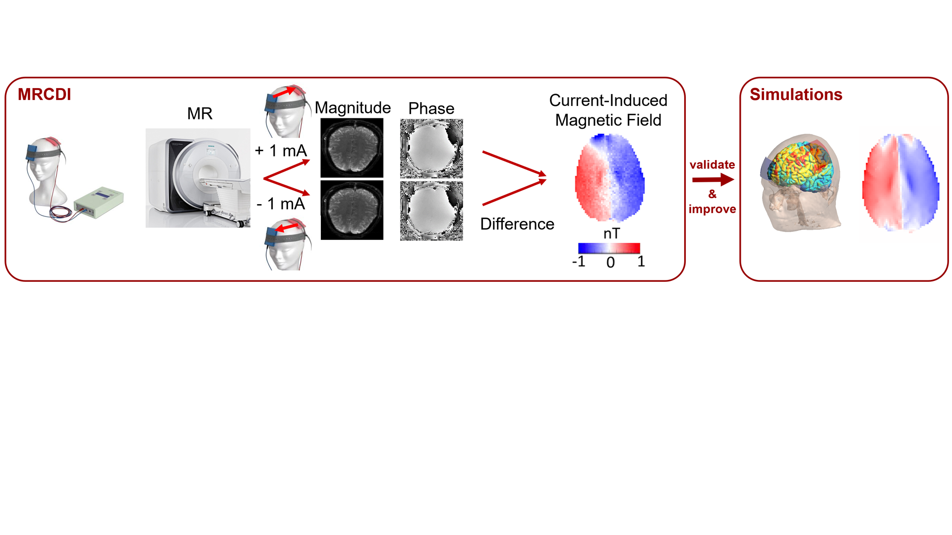
Most Transcranial Brain Stimulation (TBS) methods induce electric currents to modulate brain activity and behavior. Here, we develop and apply magnetic resonance current density imaging (MRCDI) to directly measure the current flow pattern generated by transcranial electric stimulation in the in-vivo human brain.
We have made tremendous technical advancements on MRCDI, which we now aim to apply to validate and improve simulated current flow patterns, e.g. calculated using SimNIBS (www.simnibs.org). Our hope is that this will ultimately resulting in more accurate simulations based on next-generation volume conductor models of the head. We also collaborate with MeMoSLAP to underscore how measured and simulated current flows relate to the physiological and behavioral effects of transcranial electric stimulation.
People:
Collaborators:
Group Leader
Axel Thielscher Head of Section, Professor Department of Health Technology Phone: +45 45255313 axthi@dtu.dk

